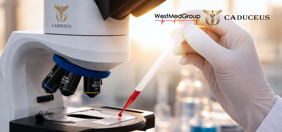
Laboratory diagnostics plays a crucial role in contemporary medicine, enabling the detection of changes in cells, tissues, and biological samples at early stages. Research accuracy depends not only on the qualifications of the specialist but also on the quality of the equipment used.
One of the key tools in this practice remains the medical microscope. Its functionality is directly determined by the capabilities of the optical system, illumination technologies, and the quality of digital visualization.

Modern models combine high image detail with ease of use. The foundation of these microscopes consists of objectives, which form the primary image of the sample under study and determine the level of detail.
For example, CADUCEUS RESEARCH BS microscopes use achromatic objectives with magnifications of 4x, 10x, 40x, and 100x, providing basic correction of chromatic aberrations. This reduces color distortion and enhances overall image clarity. The high resolution of such optics enables detailed study of various tissues and microorganisms.

Additionally, the visual observation system plays a significant role. Wide-field eyepieces with diopter adjustment increase comfort during prolonged use and reduce eye strain. A trinocular design allows for simultaneous visual observation and the connection of a digital camera to display the image on a monitor, capture photos and videos, or conduct collaborative analysis of the material.
Different microscopy methods are used depending on the task:
Thus, modern medical microscopes combine various optical solutions, illumination systems, and contrast methods. The technological differences between models are determined by the level of optical correction, image formation quality, and the range of available microscopy modes.