Size: А А А Interval: AB AB AB Colors: А А А А Images: Включить графику Выключить графику
Regular versionRegular version

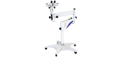
Slit lamp CADUCEUS EYESLIT MT

Slit lamp CADUCEUS MT

is a device consisting of a narrow-beam light source and a binocular microscope used for biomicroscopic analysis of the eye’s visible parts: eyelids, sclera, conjunctiva, iris, lens and cornea. The researcher can avoid connecting external photo and video fixation devices, providing convenience and integration into the diagnostic process. A slit lamp allows to examine the structures of the eye in side lighting, which helps to identify various pathologies and assess the condition of tissues. Slit lamps are used to diagnose and monitor the treatment of diseases in ophthalmology, such as cataracts, glaucoma, corneal dystrophy, keratitis and others.

Advantages:

Easy to use and requires minimal training to perform the inspection
The ability to examine tissues improves the accuracy of diagnosis and the effectiveness of treatment of eye diseases
Designed to meet safety and hygiene standards, which ensures the protection of the health of patients and staff
The devices have a long service life and require minimal maintenance

Specifications

Magnification change drum 5-steps of magnification
Eyepiece magnification, x 12,5Х
Total magnification, x 6Х; 10Х; 16Х; 25Х
Working distance, ±10%, mm 100
Field of view diameter, ±10% mm 33; 22,5; 14; 8,8
Eyepiece adjustment, dptr from -7 to +7
Interpupillary distance adjustment, ±10%, mm from 52 to 82
Filters heat-absorbing,
gray,
no red,
blue 2 transparent,
green,
blue
Slit image width, ±10%, mm smooth adjustment
from 0 to 14
Aperture, ±10%, mm 14; 10; 5; 2; 1; 0,2
Slot angle, ° -90/0/90
Carriage (movable base) movement:  
– Horizontal left-right, ±10%, mm 110
– Horizontal forward-backward, ±10%, mm 115
Vertical movement of the face stop, ±10%, mm 80
Lighting type Upper
Lighting source LED
Lighting power supply DC current, 5V 3A
Maximum gap brightness, lux No less then 50 000
Brightness adjustment smooth
Camera type Built-in digital camera (without external photo attachment)
Shooting modes Static pictures (photos)
Dynamic recording (video)
Shooting Settings Aperture (AV)
Shutter speed (TV)
Sensitivity (ISO)
Exposure (BB)
Save formats Photo: JPEG, PNG
Video: AVI or MP4
Image labeling Automatic Left (L) / Right (R) Eye Marking
Scaling Programmatically enlarge images in the interface
Connection USB Data Cable
Video recording Real-time recording with timestamps
Playback via interface
Illumination Dimmable LED source
Filters: Heat Absorbing, Grey, Redless, Cobalt Blue
Additional features Diagnostic templates for quick conclusion
Search for patients by keywords

* Specifications are subject to change without notice.

Documents

You can request additional information about the equipment, as well as find out the cost and place an order by writing to us at info@westmedgroup.ru
Centered telescopic optical system with automatic lens type detection and measurement.
An ophthalmic device for checking, measuring, training and improving the patient's binocular vision, as well as eliminating disorders of an eye motor functions.
An optical instrument with a wide range of magnifications and focal lengths for accurate diagnosis and effective treatment of eye diseases.
Medical laboratory microscope for various histological and microbiological studies
Laboratory medical microscope